

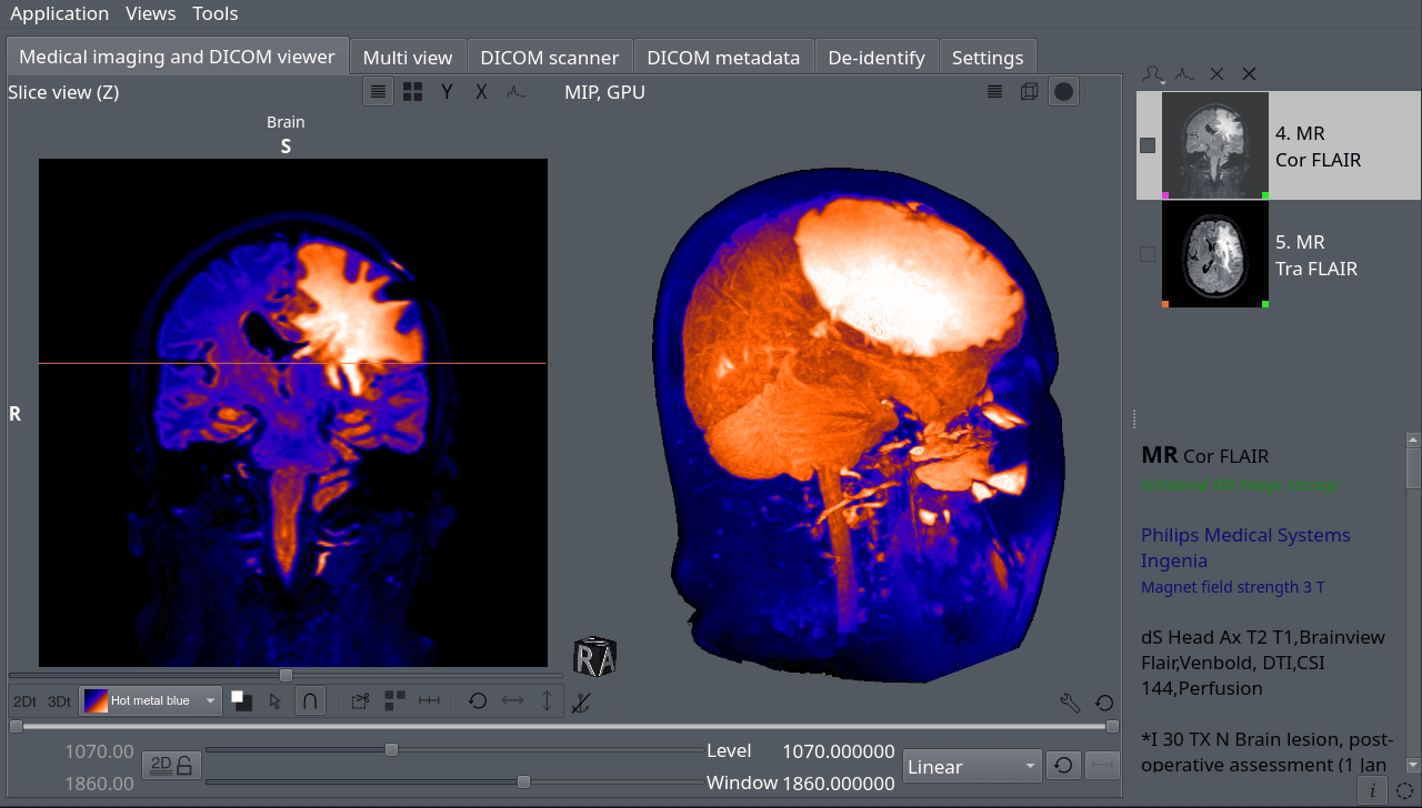
Aliza MS is a medical imaging and DICOM viewer designed for examining clinical imaging studies on the desktop.
Its workflow centres on a DICOM scanner: users can open a directory containing DICOM files or a DICOMDIR file, select one or more series, and load them into the viewer. Directories can also be opened with drag-and-drop. It supports both conventional and time-resolved datasets, with tools for navigating and analysing studies in multiple dimensions. The project provides native Linux builds and can also be built from source on Linux.
This is free and open source software.
Key Features
- 2D and 3D views with a broad collection of viewing and manipulation tools.
- 3D viewing of non-uniform image series.
- Volume rendering for visualising volumetric datasets.
- Maximum intensity projection for highlighting high-intensity structures.
- Multi-planar reconstruction for viewing image data along different planes.
- Fast directory scanning together with DICOMDIR support.
- Consistent DICOM de-identification while maintaining study integrity.
- RTSTRUCT contour display for radiotherapy structure sets.
- Multi-view display of DICOM studies with intersection lines and side-by-side views.
- Measurements within Ultrasound Calibrated Regions.
- 2D+t and 3D+t animation for time-resolved imaging datasets.
- DICOM metadata viewer using the DICOM 2026b dictionary.
- Support for encapsulated transfer syntaxes, including High-Throughput JPEG 2000.
- Support for most DICOM Information Object Definitions, including Structured Reports, Key Object Selection and Grayscale Softcopy Presentation State.
- Keyboard shortcuts for fitting images, toggling smoothing and displaying image information.
- Screenshot capture directly from 2D and 3D views.
- Optional linear filtering for the 3D view.
- Frame display for inspecting non-uniform images in 3D physical space.
Website: github.com/AlizaMedicalImaging/AlizaMS
Support:
Developer: Aliza Medical Imaging
License: GNU General Public License v3.0
Aliza MS is written in C++ and C. Learn C++ with our recommended free books and free tutorials. Learn C with our recommended free books and free tutorials.
Related Software
| Medical Imaging Software | |
|---|---|
| 3D Slicer | General purpose biomedical software with visualization and analysis |
| XMedcon | Medical Image Conversion Utility |
| Weasis | Multipurpose standalone and web-based DICOM viewer |
| OHIF Viewer | Browser-based medical imaging viewer |
| MRIcroGL | Provides tools for 2D and 3D display of medical images |
| GATE | Numerical simulations in medical imaging and radiotherapy |
| dcm4che | DICOM implementation in Java |
| AMIDE | A Medical Imaging Data Examiner |
| InVesalius | Generate 3D medical imaging reconstructions |
| SMILI | The Simple Medical Imaging Library Interface |
| ITK-SNAP | Multi-platform medical image segmentation application |
| ImageJ | General image processing program with DICOM support |
| MITK | Medical Imaging Interaction Toolkit |
| Starviewer | Medical imaging software fully compliant with the DICOM standard |
| Orthanc | Lightweight DICOM server |
| DicomBrowser | Inspect and edit DICOM header fields, and DICOM viewer |
| Aeskulap | Medical image viewer |
| openDICOM.NET | DICOM library project |
| kradview | DICOM viewer |
| Ginkgo CADx | Advanced DICOM viewer and dicomizer with conversion functionality |
Read our verdict in the software roundup.
 Explore our carefully curated directory of recommended free and open source software, covering every major software category. Explore our carefully curated directory of recommended free and open source software, covering every major software category.The directory forms part of our extensive collection of articles for Linux enthusiasts. It includes hundreds of detailed reviews, together with free and open source alternatives to proprietary software from companies such as Google, Microsoft, Apple, Adobe, IBM, Cisco, Oracle, and Autodesk. LinuxLinks also covers interesting projects worth exploring, Linux-compatible hardware, free programming books and tutorials, and much more. Know a useful free and open source Linux application that we haven’t covered? Tell us about it using our submission form. |
Please read our Comment Policy before commenting.